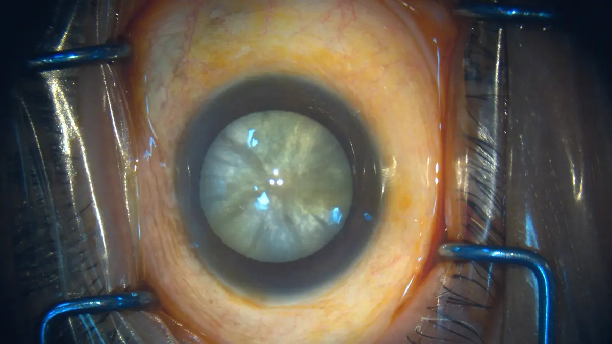
Dense, white or mature cataract
Reduced red reflex and a hard nucleus may require modified capsule techniques and careful energy management.


Anticipate • Prepare • Protect • Delhi Eye Care
Dense cataracts, weak lens support, small pupils and coexisting retinal, corneal or glaucoma disease require additional examination, preparation and counselling.
Complexity factors
Hard nuclei may require greater energy and careful protection of the cornea.
Pseudoexfoliation, trauma or previous surgery may reduce support for the lens capsule.
Medication, scarring or pseudoexfoliation may require pupil-expansion strategies.
Macular status affects visual potential and may alter treatment timing.
Pressure control, medication and possible combined procedures need consideration.
Endothelial health and irregular astigmatism can influence technique and recovery.
Joined-up eye care
A technically successful cataract operation cannot reverse vision loss caused by advanced macular, optic-nerve or corneal disease. Where required, DEC coordinates cataract planning with retina, glaucoma or cornea assessment.
Dr. Nidhi may adapt the incision, anaesthesia, pupil management, lens choice or postoperative schedule. Additional devices or a staged approach may occasionally be appropriate.
First, a reassuring distinction
The surgeon anticipates factors that may make access, cataract removal, lens support or recovery less predictable than in a routine eye.
An unintended problem occurring during or after surgery. Recognising complexity beforehand helps the team prepare to reduce and manage risk.
Patterns that change the plan
Reduced red reflex and a hard nucleus may require modified capsule techniques and careful energy management.
Pseudoexfoliation, trauma, high myopia, previous surgery or inherited conditions may weaken zonules.
The pupil may not dilate adequately because of medicines, pseudoexfoliation, inflammation or previous procedures.
Low endothelial reserve, guttata, scars or irregular shape can influence energy use, incision and recovery expectations.
Pressure control, filtering blebs, narrow angles and optic-nerve reserve affect timing and postoperative monitoring.
Diabetic retinopathy, AMD, epiretinal membrane, previous detachment or vitrectomy can limit vision and change coordination.
Corneal refractive surgery, vitrectomy, injections, trauma or inflammation can affect measurements and anatomy.
Tremor, breathing difficulty, inability to lie flat, anticoagulants and cooperation influence anaesthesia and theatre planning.
Short or long eyes and previous LASIK/PRK require careful biometry, formula selection and expectation setting.
Plan before entering theatre
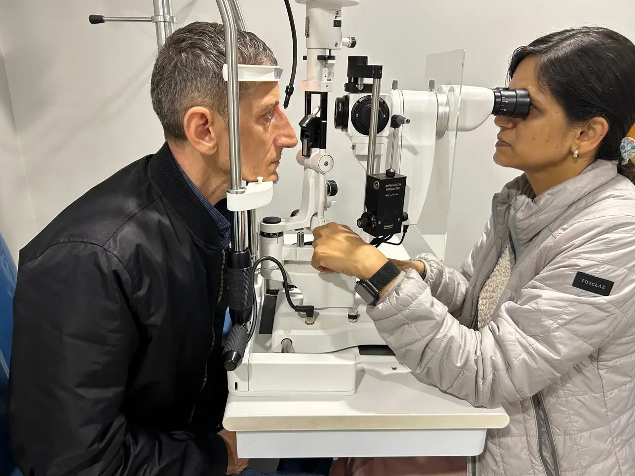
Slit-lamp examination, pupil dilation, eye pressure and dilated fundus evaluation identify the main challenges.
OCT, corneal testing or ultrasound may be used when the cataract or coexisting disease obscures the prognosis.
Dr. Nidhi plans the preferred method as well as devices, lens options and contingencies that may become necessary.
The review schedule and medicines are adjusted to corneal, retinal, pressure or inflammatory risk.
Surgical strategies
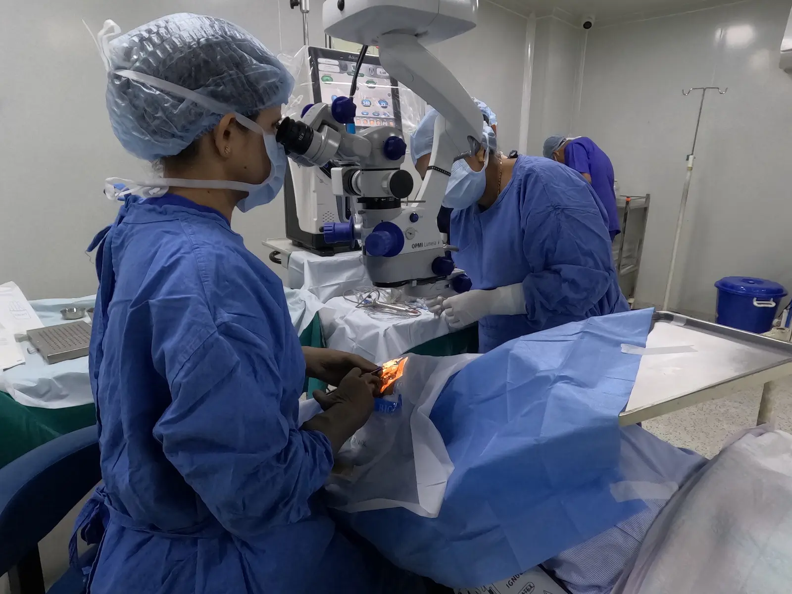
A complex plan may include one or more supportive measures. Their use is determined intraoperatively as well as from preoperative findings.
Mechanical devices may improve safe visibility when pharmacological dilation is inadequate.
Rings, hooks or other supports may stabilise a capsule affected by weak zonules.
Dense nuclei may require changes in fragmentation, fluidics and corneal protection.
If capsular support is insufficient, the safest lens position or staged option may differ from the original preference.
Coexisting retinal disease
| Finding | Why it matters | Possible planning response |
|---|---|---|
| Diabetic macular oedema or active retinopathy | May limit central vision or worsen around surgery. | Retinal assessment and treatment may be coordinated before, around or after cataract surgery. |
| AMD or macular scar | Cataract removal can improve clarity but cannot reverse macular damage. | OCT and realistic visual-potential counselling guide timing and IOL choice. |
| Previous retinal detachment or vitrectomy | Anatomy, lens support and retinal risk may differ. | Detailed peripheral retinal review and modified surgical planning may be appropriate. |
| Epiretinal membrane or macular hole | Distortion or central blur may persist after cataract removal. | Retina and cataract sequencing is individualised. |
Led by Dr. Nidhi Gupta
Dr. Nidhi Gupta leads cataract care at Delhi Eye Care with over 18 years of experience and 15,000+ ophthalmic surgeries. Her approach is to identify risk, explain what it changes and prepare alternatives without creating unnecessary anxiety.
“Complex does not mean hopeless. It means that the eye deserves a more deliberate plan, realistic expectations and the right support around surgery.”
Patients and families are told what is known before surgery, what may only become clear during the operation, and how the postoperative schedule may differ.
Setting the right expectation
Whether the cloudy lens can be removed and an IOL supported safely.
How the cornea, inflammation and eye pressure are expected to settle.
What the retina, macula and optic nerve are capable of providing after the media becomes clear.
When to seek early review after surgery
Contact Delhi Eye Care promptly for increasing pain, marked redness, sudden worsening of vision, new flashes or floaters, a curtain-like shadow, discharge, nausea with eye pain, or any concern that recovery is moving backwards rather than forwards.
Common questions
No. It means additional factors may increase technical difficulty or affect recovery and visual potential. Identifying them allows more careful planning and counselling.
Usually yes, but a dense cataract may require modified techniques, more energy and additional precautions. Examination and, when the retina cannot be seen, ultrasound may be required.
Zonules are fine fibres supporting the natural lens capsule. Pseudoexfoliation, trauma, previous surgery and other conditions can weaken them, sometimes requiring capsular-support devices or a different IOL plan.
It may improve the component of blur caused by cataract, but it cannot reverse retinal damage. OCT and retinal assessment help estimate realistic visual potential.
Not always. Lens choice depends on measurements, corneal and retinal health, capsular support and visual priorities. In some eyes, optical simplicity and secure positioning are more important than a wider range of focus.
Corneal swelling, pressure, inflammation, retinal status or lens support may need closer monitoring than after an uncomplicated routine case.
Continue exploring
Delhi Eye Care
A careful evaluation helps separate what is possible from what is appropriate for your eye.
Call: +91 92208 92424
Specialist: Dr. Nidhi Gupta, Medical Director
Cataract, Refractive & Glaucoma Surgeon
Medically reviewed by Dr. Nidhi Gupta • Last reviewed September 2026