01
Eye pressure
Is the pressure safe for your optic nerve?


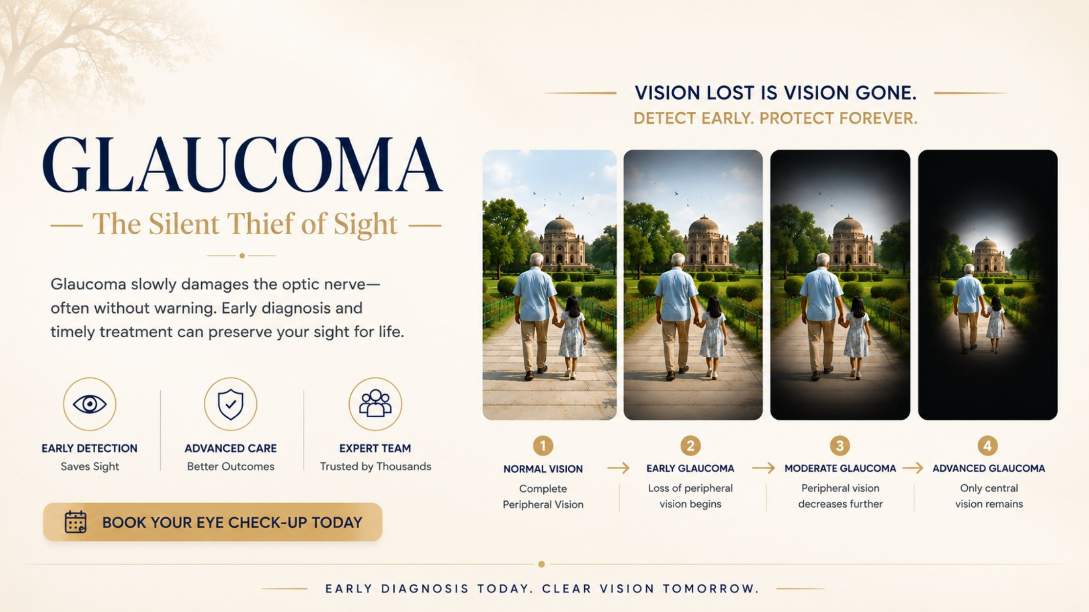
Glaucoma can damage the optic nerve silently. Our care is built around accurate diagnosis, an individual target eye pressure, and long-term monitoring of both the optic nerve and visual field.
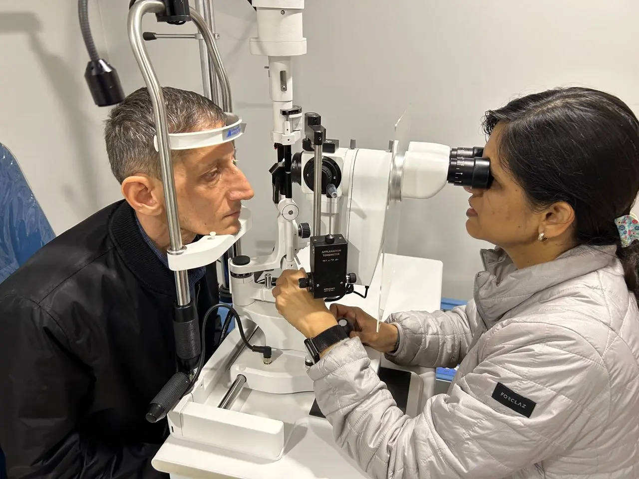
A single pressure reading is only one piece of the assessment. Diagnosis and treatment decisions come from the relationship between pressure, anatomy, nerve health, visual function and progression.
Is the pressure safe for your optic nerve?
Is there evidence of glaucomatous damage?
Is the angle open, narrow or closed?
Has glaucoma affected functional vision?
Is the disease stable—or changing over time?
A pressure of 18 may be safe for one eye and unsafe for another. We treat the patient and the optic nerve—not simply a number on the tonometer.
Choose the situation closest to yours. The examination is then planned around the question that needs answering.
You may have ocular hypertension without established glaucoma. We assess whether the optic nerve is genuinely at risk.
Understand the next stepA suspicious disc, family history or borderline test does not always mean established glaucoma.
Build a reliable baselineUnderstand the type, stage, target pressure and whether your disease is stable.
Plan long-term controlNarrow or closed angles require a different strategy from open-angle glaucoma.
See glaucoma typesTreatment may be cataract surgery alone, staged treatment, or combined surgery.
Explore the optionsThe diagnosis, target pressure, adherence, laser and surgical options may need to be reconsidered.
Seek a second opinionWe bring clinical examination, structural imaging and functional testing together to decide whether glaucoma is present, how advanced it is and whether it is changing.
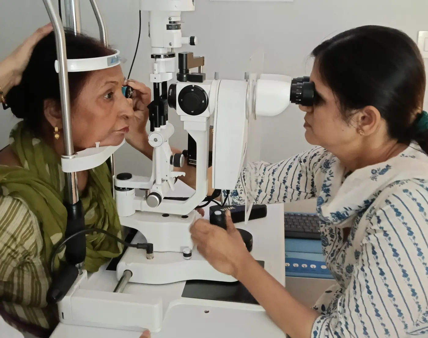
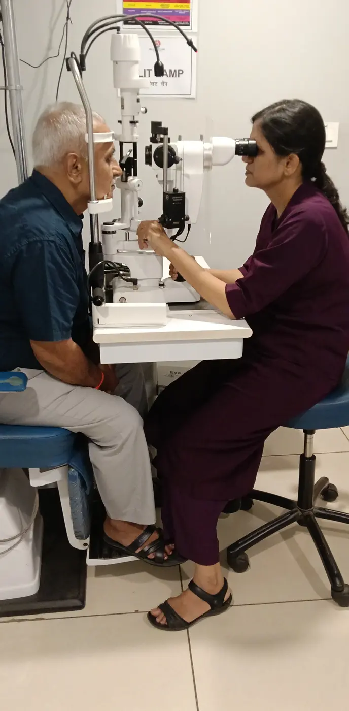
Direct examination of the drainage angle to distinguish open-angle from angle-closure disease.
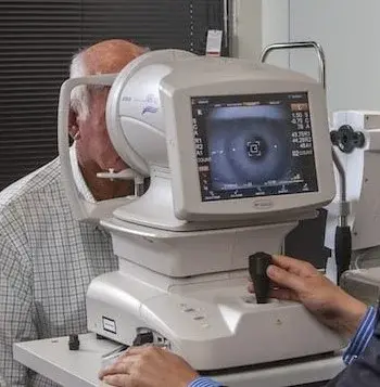
RNFL and ganglion-cell analysis assess glaucoma-related structural loss.
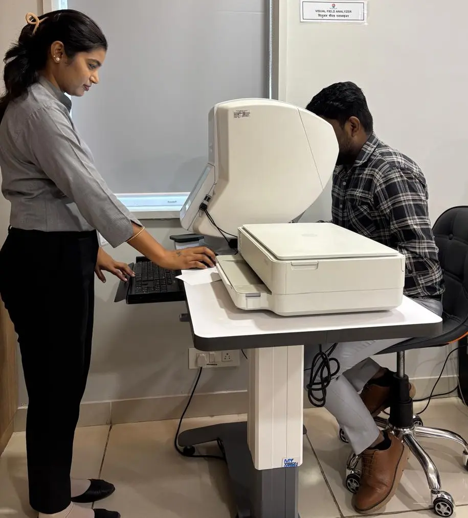
Measures functional impact and helps identify meaningful progression.
Accurate measurement of intraocular pressure.
Pachymetry helps place the measured pressure into its proper clinical context.
Clinical assessment and documentation for comparison over time.
Its type, severity, angle anatomy, rate of change and the needs of the individual eye all influence treatment.
The common chronic form; usually develops without early symptoms.
Optic-nerve damage can occur even when pressure is within the statistical normal range.
A narrow or closed drainage angle may require laser, lens-based treatment or surgery.
Not everyone with raised pressure has glaucoma—and not every glaucoma patient has raised pressure.
Steroid-related, uveitic, neovascular, pseudoexfoliative, pigmentary and other secondary forms.
Eyes needing very low targets, surgery, care after prior surgery or management with other eye disease.
Treatment begins with the target pressure and risk of future visual loss. These are individual options—not a fixed treatment ladder.
For selected suspects and lower-risk patients with a reliable baseline.
Selective laser treatment may be considered early or later for suitable open-angle disease.
Chosen according to target pressure, effectiveness, tolerability and general health.
For suitable patients within the narrow-angle and angle-closure spectrum.
Particularly relevant in selected patients with cataract and crowded anterior segments.
When adequate control cannot be achieved safely with less invasive treatment.
Having cataract and glaucoma does not automatically mean that both require surgery at the same time. The right sequence depends on glaucoma type, stage, target pressure, angle anatomy and the visual effect of the cataract.
The aim is not simply to remove the cataract or lower pressure. It is to choose the strategy that gives the eye the safest long-term visual future.
Glaucoma care is a continuing relationship. Tests are repeated at intervals suited to disease stage, risk and evidence of change. With timely diagnosis, appropriate treatment and regular follow-up, most people maintain useful vision and a good quality of life.

Cataract, Refractive & Glaucoma Surgeon with dedicated experience in medical and surgical glaucoma care, complex cataract surgery and eyes where cataract and glaucoma occur together.
GSI · AIOS · DOS · NZOS · HOS
If a parent, brother, sister or child has glaucoma, tell your ophthalmologist. Close family members should have a comprehensive eye examination and an appropriate screening schedule.
Plan a family eye checkA specialist review can help when the pressure remains above target or the disease appears to be progressing.
Bring previous fields, OCT scans, prescriptions and operative records whenever possible.
This may be an acute angle-closure attack. Call immediately. If DEC cannot be reached promptly, attend the nearest emergency eye service—do not wait for a WhatsApp response.
If you have raised pressure, a family history, an abnormal optic nerve or an established diagnosis, let us determine whether your vision is at risk and what needs to be done.
Medically reviewed by Dr. Nidhi Gupta · October 2026
This page is for general education and does not replace an individual medical examination.