Delhi Eye Care Retina Excellence Centre Elementor integration block.
Delhi Eye Care
Retina Excellence CentreClarity at every step of
retina care.
Comprehensive medical and surgical retina care in Delhi, led by
senior vitreo-retina surgeon
Dr Shashank Rai Gupta—from detailed imaging and
careful monitoring to laser, injections and complex vitreoretinal
surgery.
The retina is small. Its role in sight is immense.
The retina is the light-sensitive tissue at the back of your eye.
Some retinal conditions cause obvious visual symptoms; others
progress quietly. A detailed retinal examination and appropriate
imaging help us understand what is happening—and whether the
safest next step is observation, treatment or surgery.
I have new symptoms
Identify what the change may mean and how urgently you should
seek a retinal assessment.
Floaters may look like dots, threads, cobwebs, rings or a moving
shadow. Flashes may appear as brief sparks or arcs, often at the
side of vision. Many come from age-related change in the clear
vitreous gel, but a sudden change can be the first sign of a retinal
tear or detachment.
What floaters and flashes may look likeThis is a visual simulation. Actual symptoms vary: floaters
usually move with the eye, while flashes may be brief and more
noticeable in dim light or at the edge of vision.
New floaters or flashes?
The pattern and timing matter more than the presence of a floater
alone.
Often less concerning
A few familiar floaters present for months or years
No sudden increase, flashes, shadow or loss of vision
Symptoms that remain stable in both number and appearance
Arrange a prompt dilated retina examination
A sudden shower or marked increase in floaters
New flashes, a dark curtain or missing side vision
Blurred or reduced vision, especially after injury or eye
surgery
What treatment is available for floaters?
Examine, explain and observe
Common age-related or posterior vitreous detachment floaters
often become less noticeable. The first priority is to rule
out a retinal tear.
Treat the underlying cause
Bleeding, inflammation, infection or diabetic retinal
disease may need medicines, injections, retinal laser or
other cause-specific care.
Floterectomy for selected patients
Floater-only vitrectomy—sometimes called floterectomy—may be
considered when persistent vitreous opacities significantly
interfere with daily vision, after careful retinal
assessment and discussion of benefits and surgical risks.
Ordinary age-related floaters are not generally dissolved by eye
drops or tablets. Retinal laser treats a retinal tear or
diseased retina; it does not remove a routine vitreous floater.
I have a new symptom
What has changed in your vision?
Use this guide to describe the symptom clearly. It cannot diagnose
the cause, but it can help you recognise when a retinal examination
should not be delayed.
Flashes of light (photopsia)
Brief lightning streaks, sparks or camera-like flashes, usually
at the edge of vision. New flashes may occur when the vitreous
gel pulls on the retina and should be assessed, especially with
new floaters.
New onset: prompt assessmentSudden increase in floaters
A new shower of moving specks, cobwebs or dark lines may follow
vitreous separation, a retinal tear or bleeding into the
vitreous. Clinicians may see pigment cells described as “tobacco
dust.”
Sudden change: prompt assessmentA curtain, veil or fixed shadow
A dark area entering from the side—or spreading towards the
centre—can indicate an evolving retinal detachment.
Do not wait: urgent retinal assessmentLoss of side vision
Missing or narrowing peripheral vision may occur suddenly with a
localised retinal detachment or more gradually with inherited
retinal disease. The timing helps determine urgency.
Wavy or distorted vision (metamorphopsia)
Straight lines appearing bent, wavy or crooked can result from
fluid, swelling or traction at the macula, including macular
degeneration or an epiretinal membrane.
A central blind or fuzzy spot (scotoma)
A persistent dark, blurred or missing patch in the centre of
sight can interfere with reading and recognising faces and needs
macular evaluation.
Hazy or blurred central vision
Reduced sharpness may develop gradually or suddenly with macular
oedema, a macular hole, bleeding or reduced retinal circulation.
Sudden, profound painless vision loss
A marked drop in vision over seconds or hours can occur with
retinal artery or vein occlusion, major vitreous haemorrhage or
retinal detachment.
Bring the name of the condition, your prescriptions and any OCT,
fundus, angiography or ultrasound images. We review what has been
found, how urgent it is, whether it is stable or progressing, and
which choices genuinely apply to your eye.
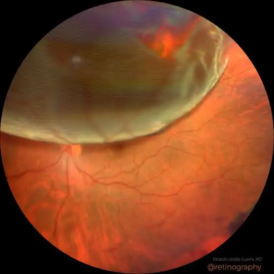
Retinal tear or detachment
Clarify the location and extent, whether the macula is
involved, and how urgently treatment is required.
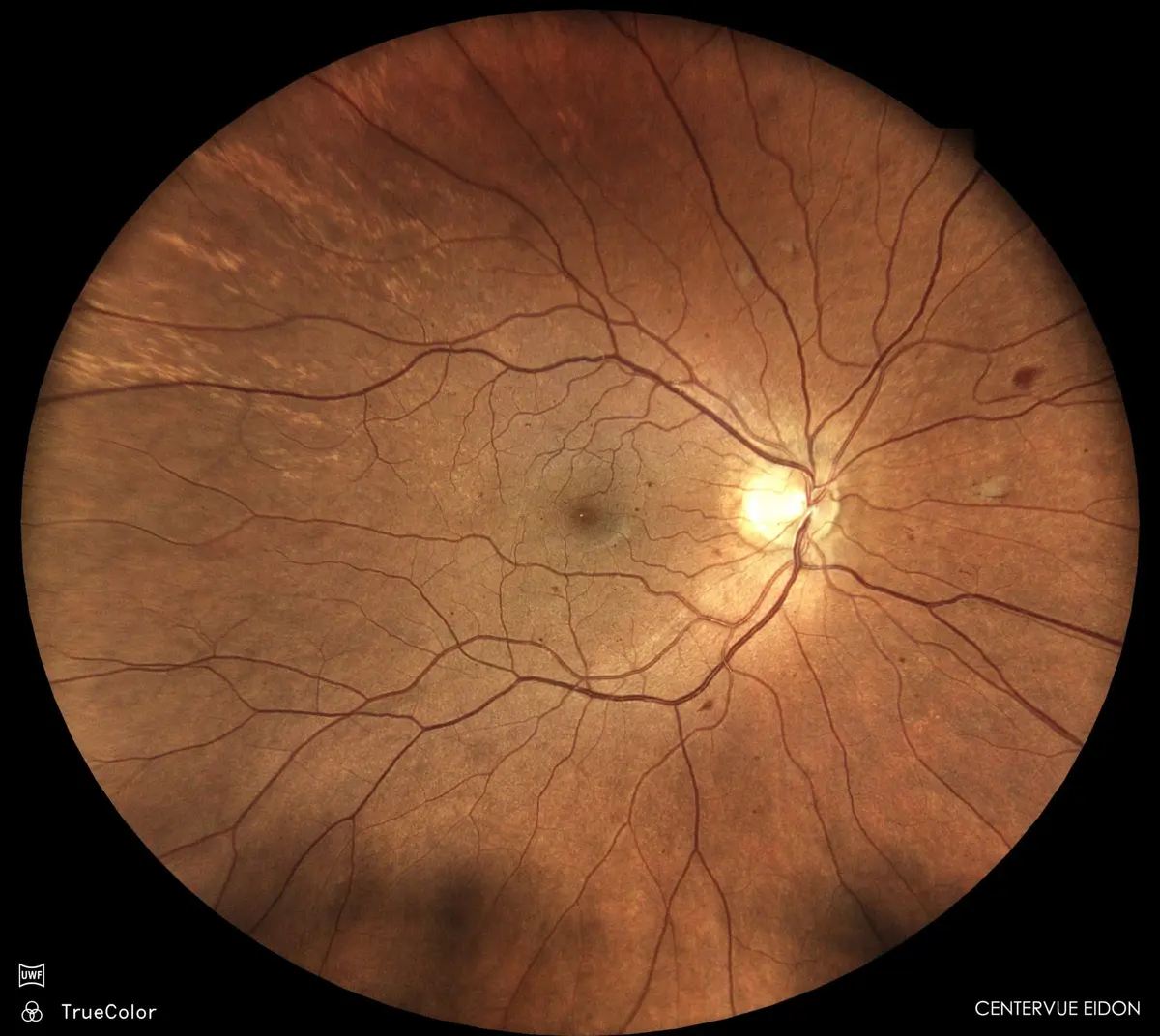
Review central retinal changes, their effect on reading or
facial recognition, and whether the macula is dry, active or
scarred.
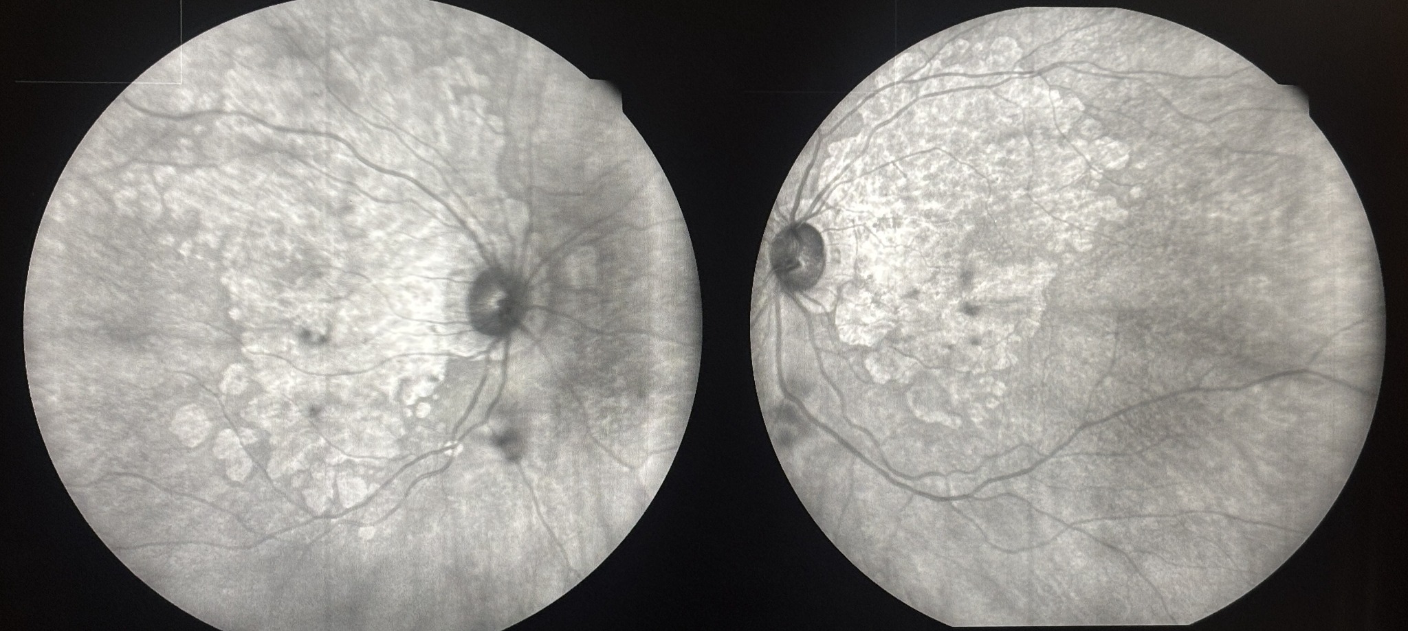
Colour retinal imagingAutofluorescence imaging
Care may include OCT-guided observation, nutritional and
lifestyle guidance for suitable dry AMD/ARMD, or intravitreal
anti-VEGF injections when neovascular activity is
present.
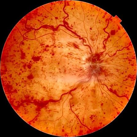
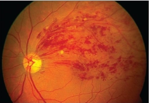
Retinal vein occlusion
Assess the distribution of bleeding, macular swelling, retinal
circulation and any abnormal new vessels.
Central retinal vein occlusion (CRVO)
Branch retinal vein occlusion (BRVO)
Treatment commonly includes OCT-guided anti-VEGF injections
for macular oedema; selected eyes may need steroid treatment,
retinal laser or monitoring for neovascular
complications.
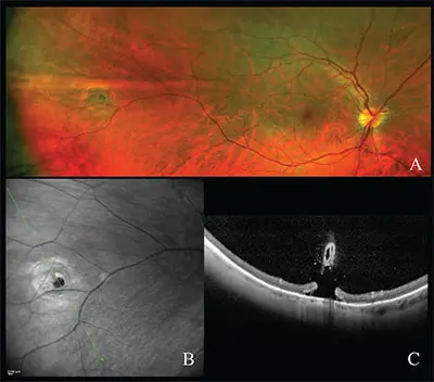
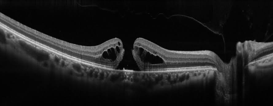
Macular hole or epiretinal membrane
Relate OCT findings to distortion, reading difficulty and the
effect on daily activities.
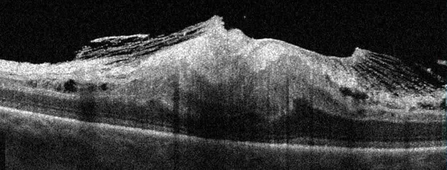
Macular hole on OCTEpiretinal membrane on OCT
Milder or stable cases may be observed. When symptoms and
anatomy justify it, vitrectomy with membrane or internal
limiting membrane peeling—and sometimes a gas bubble—may be
advised.Watch Dr Gupta discuss macular-hole surgery
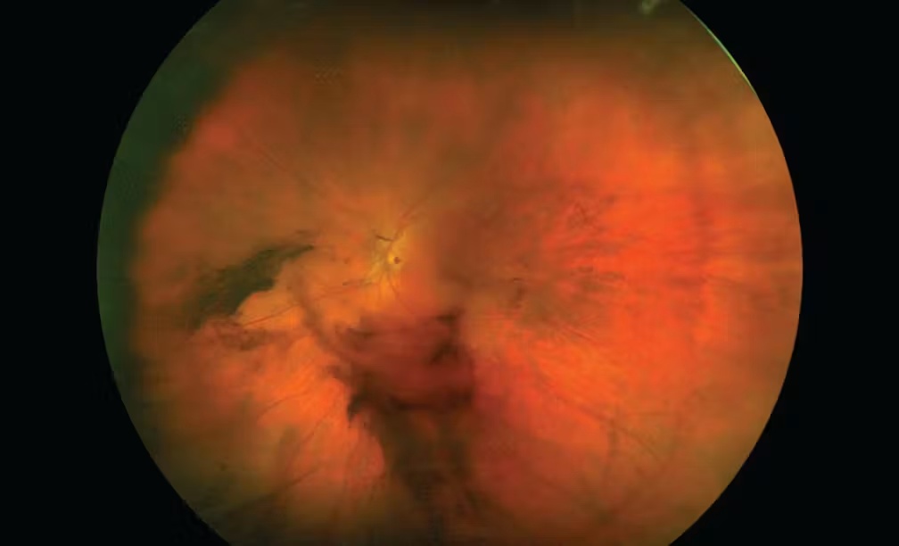
Vitreous haemorrhage or persistent floaters
Identify whether bleeding, inflammation or vitreous opacity is
responsible and rule out a retinal tear.
Vitreous haemorrhagePersistent vitreous opacity
Treatment may target the cause with injections or laser;
non-clearing haemorrhage may require vitrectomy. Floterectomy
may be considered for carefully selected, persistently
disabling floaters.Watch Dr Gupta explain floaters
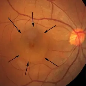
Central serous chorioretinopathy (CSCR/CSR)
Understand why fluid has collected beneath the macula, whether
this is a first episode, and if observation or treatment is
appropriate.
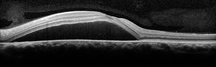
Central retinal appearanceSubretinal fluid on OCT
A recent uncomplicated episode may be monitored. Persistent,
recurrent or chronic CSCR may require multimodal imaging and
selected treatment such as photodynamic therapy or
laser.
Posterior and intermediate uveitis
Clarify whether inflammation affects the vitreous, retina or
choroid, whether infection or systemic disease is suspected,
and whether the macula or optic nerve is threatened.
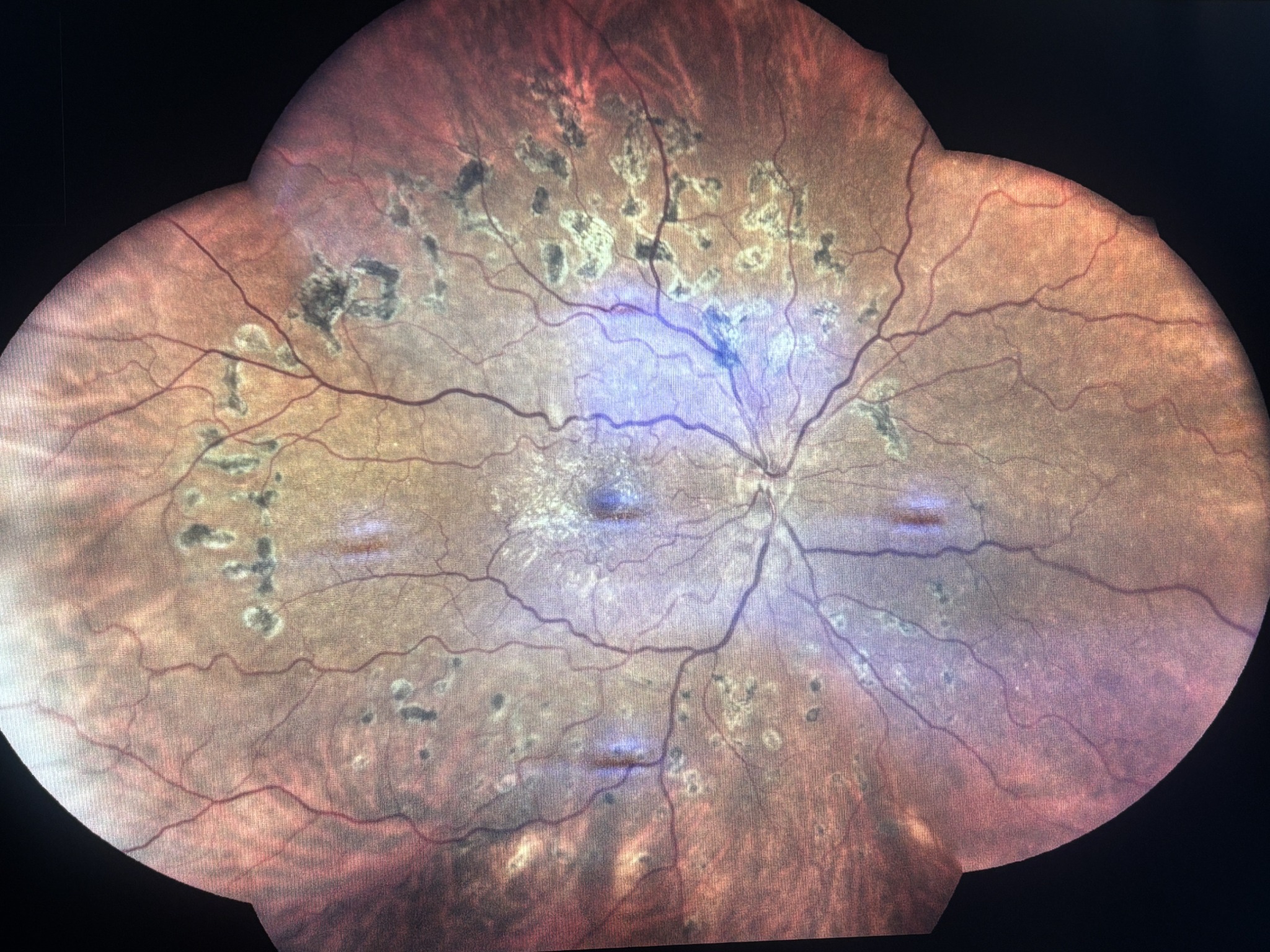
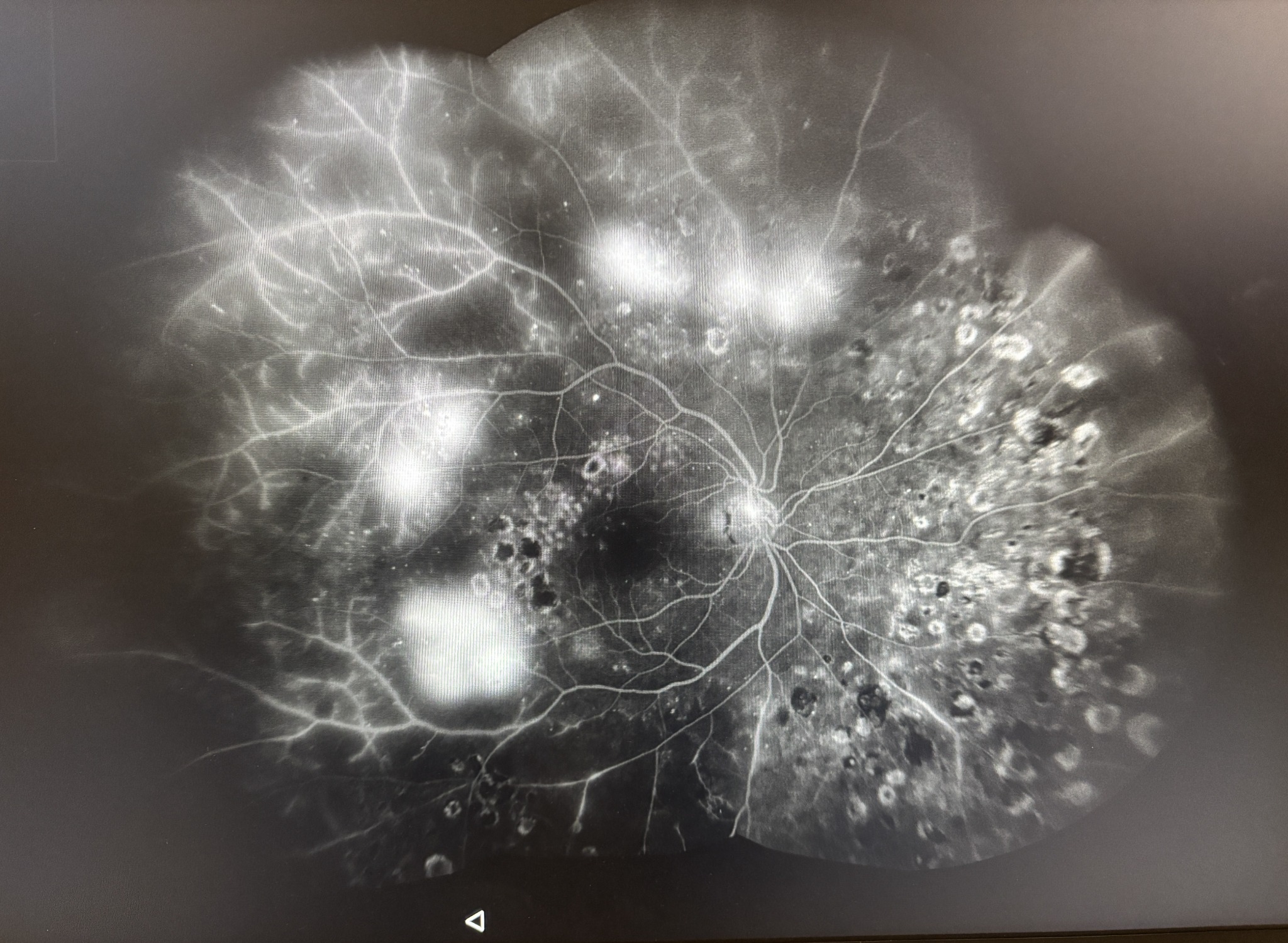
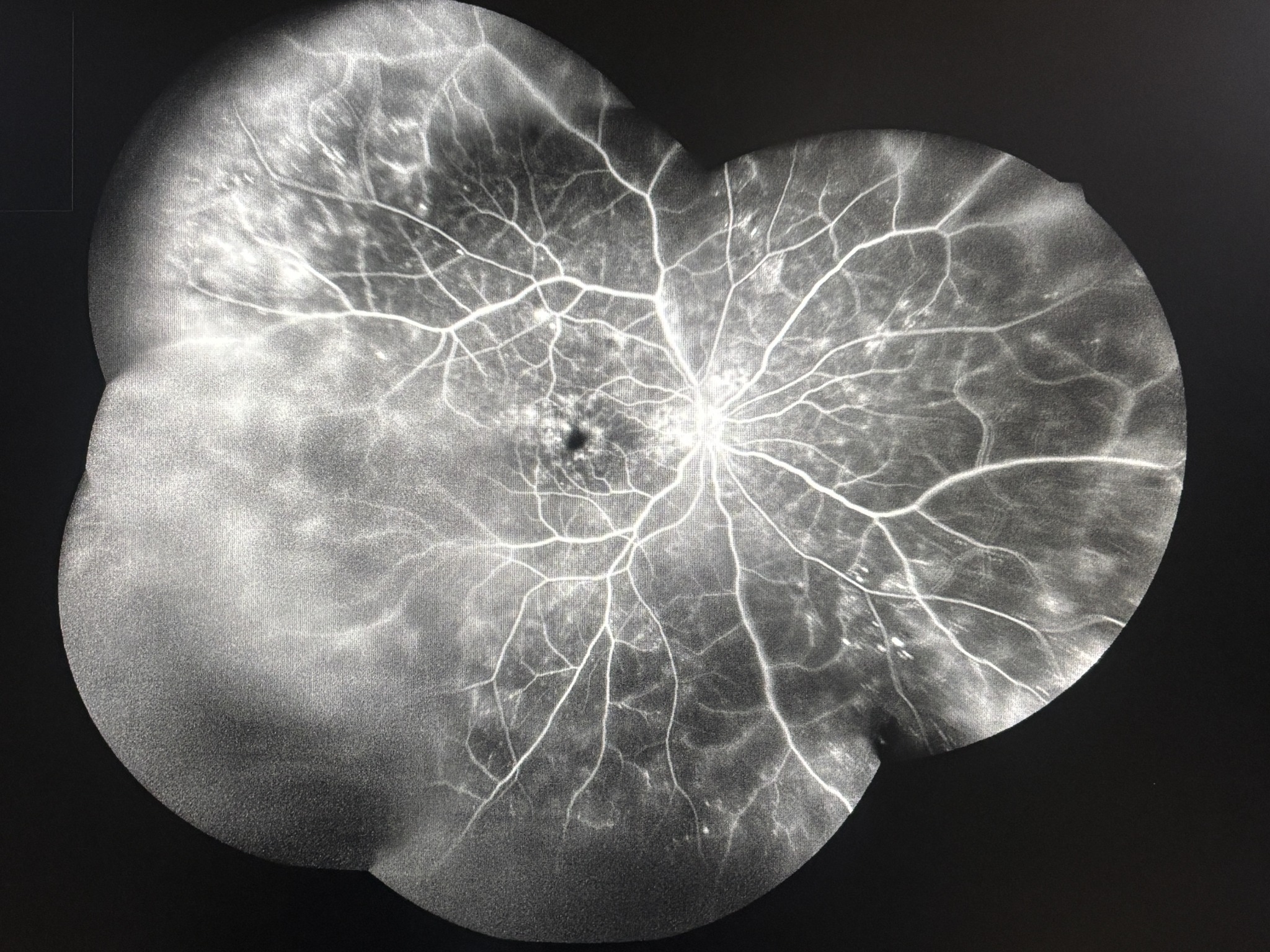
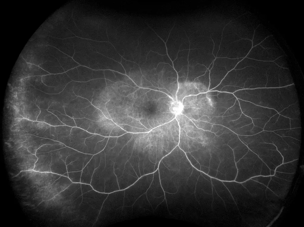
Wide-field retinal imagingFluorescein angiography
Treatment is cause-directed. Infection must be considered
before steroid or immunosuppressive treatment; selected eyes
may need local or systemic medicines, injections or
vitreoretinal surgery.
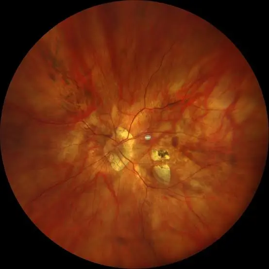
Myopia management and pathological myopia
In children, measure progression and consider evidence-based
myopia control. In high myopia, look beyond the spectacle
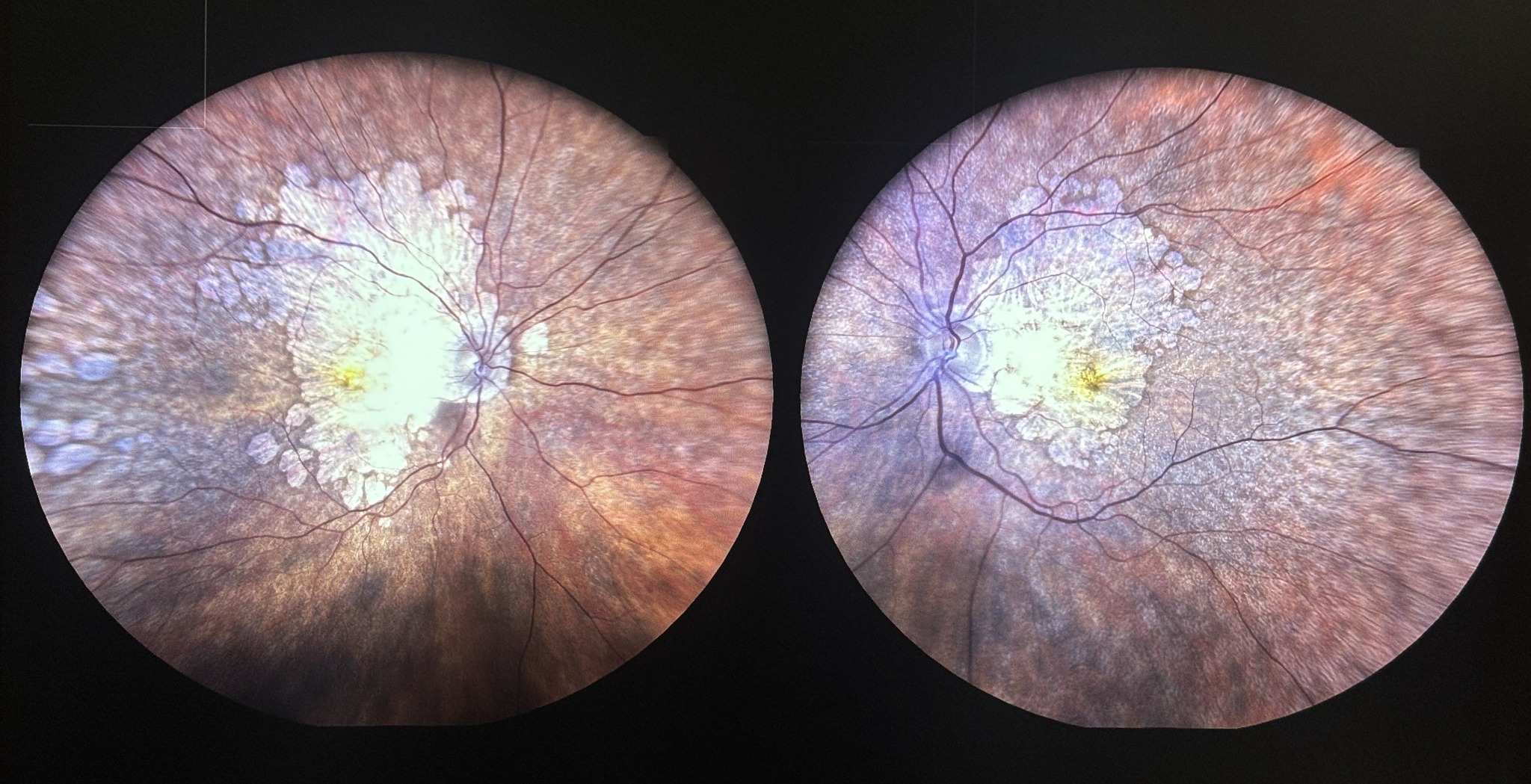
number for structural damage to the macula and retina.
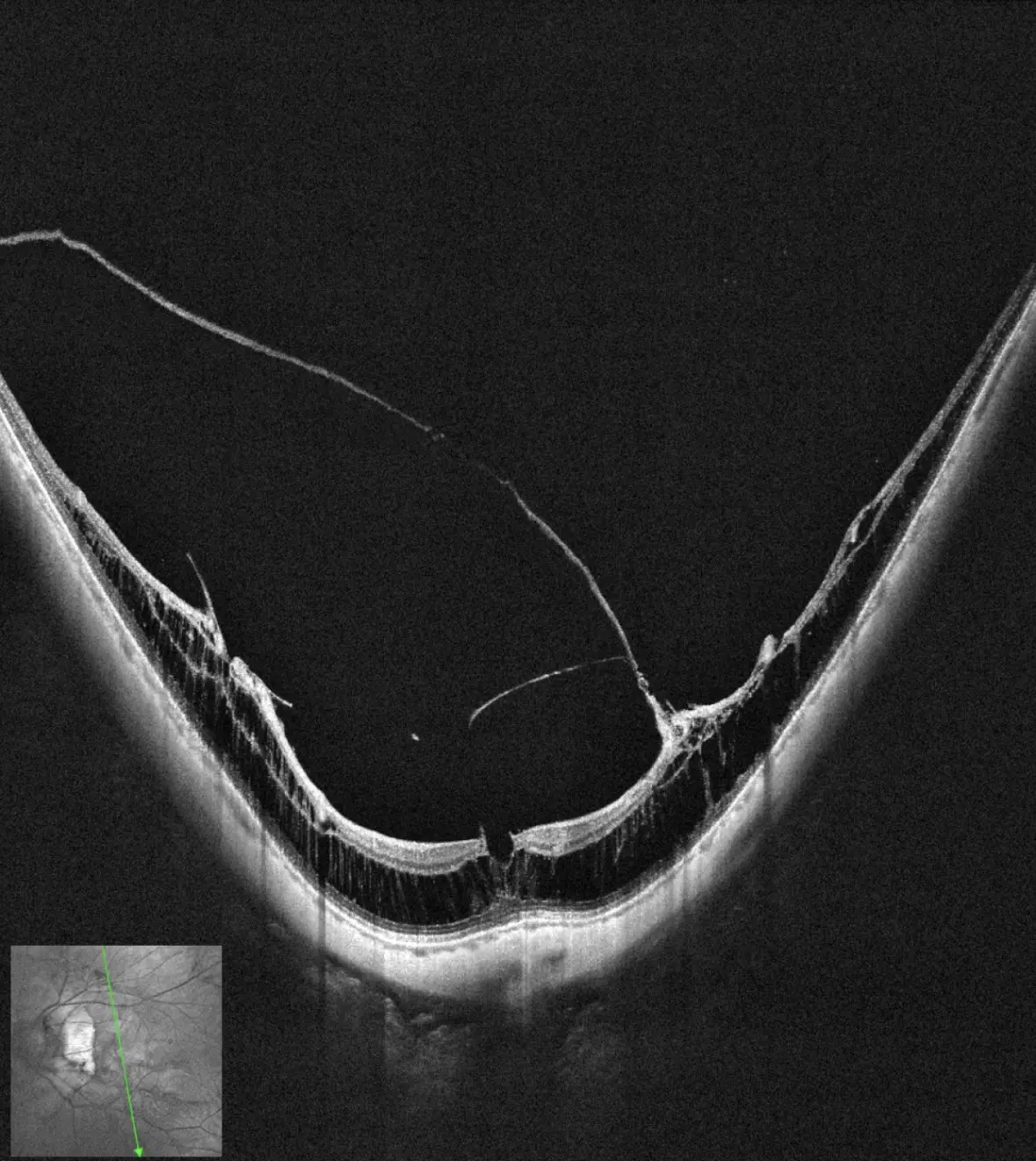
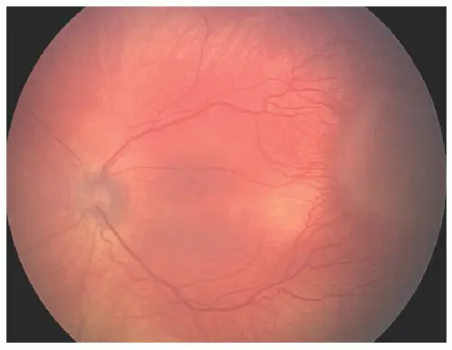
Pathological myopic retinaMyopic traction on OCT
Care may include myopia-control spectacles, contact lenses or
atropine for selected children; and monitoring, anti-VEGF,
retinal laser or surgery for specific adult retinal
complications.
Retinopathy of prematurity (ROP)
Premature babies may develop sight-threatening retinal disease
without visible symptoms. Screening and every advised review
must happen within the recommended time window.
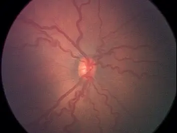
Findings are recorded by zone, stage and plus disease. Care
may include close observation, retinal laser, intravitreal
anti-VEGF or surgery for advanced tractional disease.
Other retinal conditions assessed:retinal artery occlusion, retinal trauma and
endophthalmitis. Select
a condition for its patient guide. Sudden painless vision loss and
suspected endophthalmitis require emergency assessment; retinal
artery occlusion also requires urgent systemic evaluation.
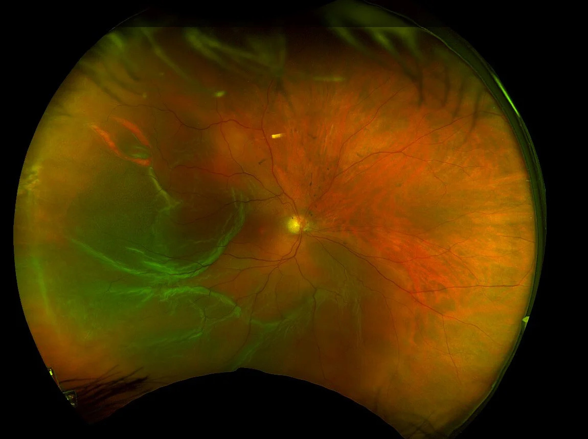
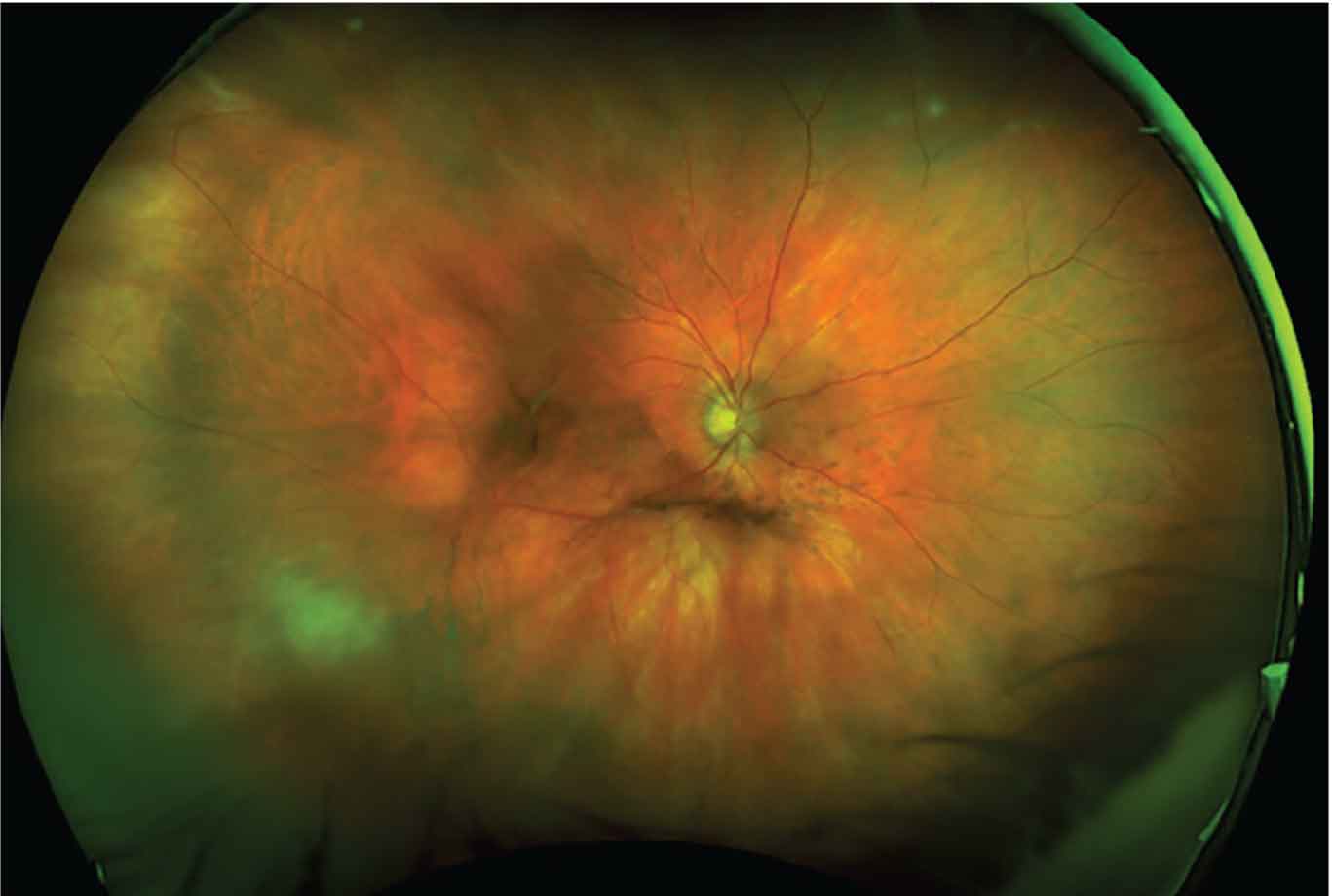
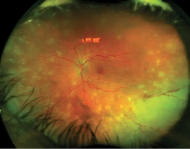
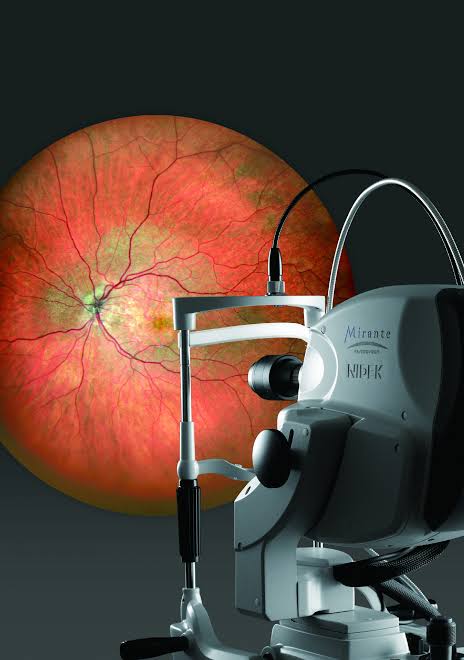
Ultra-wide-field retinal imagingDocuments a broad view of the retina, including peripheral
areas that may contain tears, vascular changes or diabetic
retinal disease.Optical coherence tomography (OCT)A rapid, non-contact scan that shows cross-sectional
retinal detail and helps assess macular fluid, holes,
membranes and treatment response.
A complete retina pathway at Delhi Eye Care
Detailed retinal examination, OCT and multimodal imaging,
injections, laser, vitreoretinal surgery and follow-up are
coordinated under Dr Shashank Rai Gupta. You receive an
explanation of the diagnosis, the available choices, the reason
for the recommendation and the expected review plan before
treatment is undertaken.
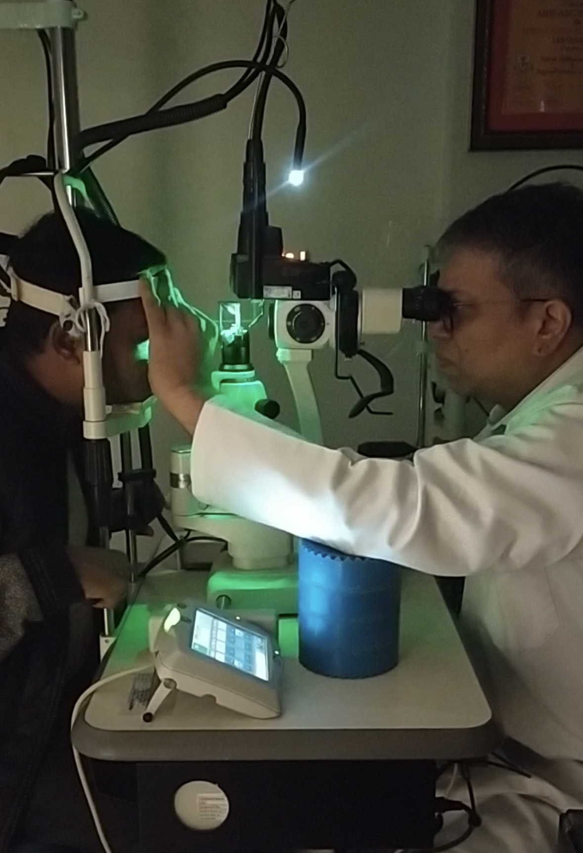
Review of symptoms, medical history and previous reports
Dilated retinal examination
OCT or other imaging when clinically needed
A clear explanation of urgency, options and follow-up
Bring previous prescriptions, scans and a medicine list. Dilation
may temporarily blur near vision and increase light sensitivity;
sunglasses and an accompanying person may help.
Not every retinal condition needs intervention. When treatment is
required, your options and likely follow-up are explained before a
decision is made.
Monitor & protect
Planned observation, serial imaging, systemic risk-factor
coordination and structured review.
Treat without surgery
Intravitreal injections, retinal laser, cryotherapy and
condition-specific medical treatment.
Vitreo-retinal surgery
Vitrectomy and appropriate retinal surgery for detachment,
macular disorders, vitreous haemorrhage and complex diabetic
disease.
Green retinal laserLaser treatment may be used to seal suitable retinal tears or
treat selected vascular and diabetic retinal conditions. The
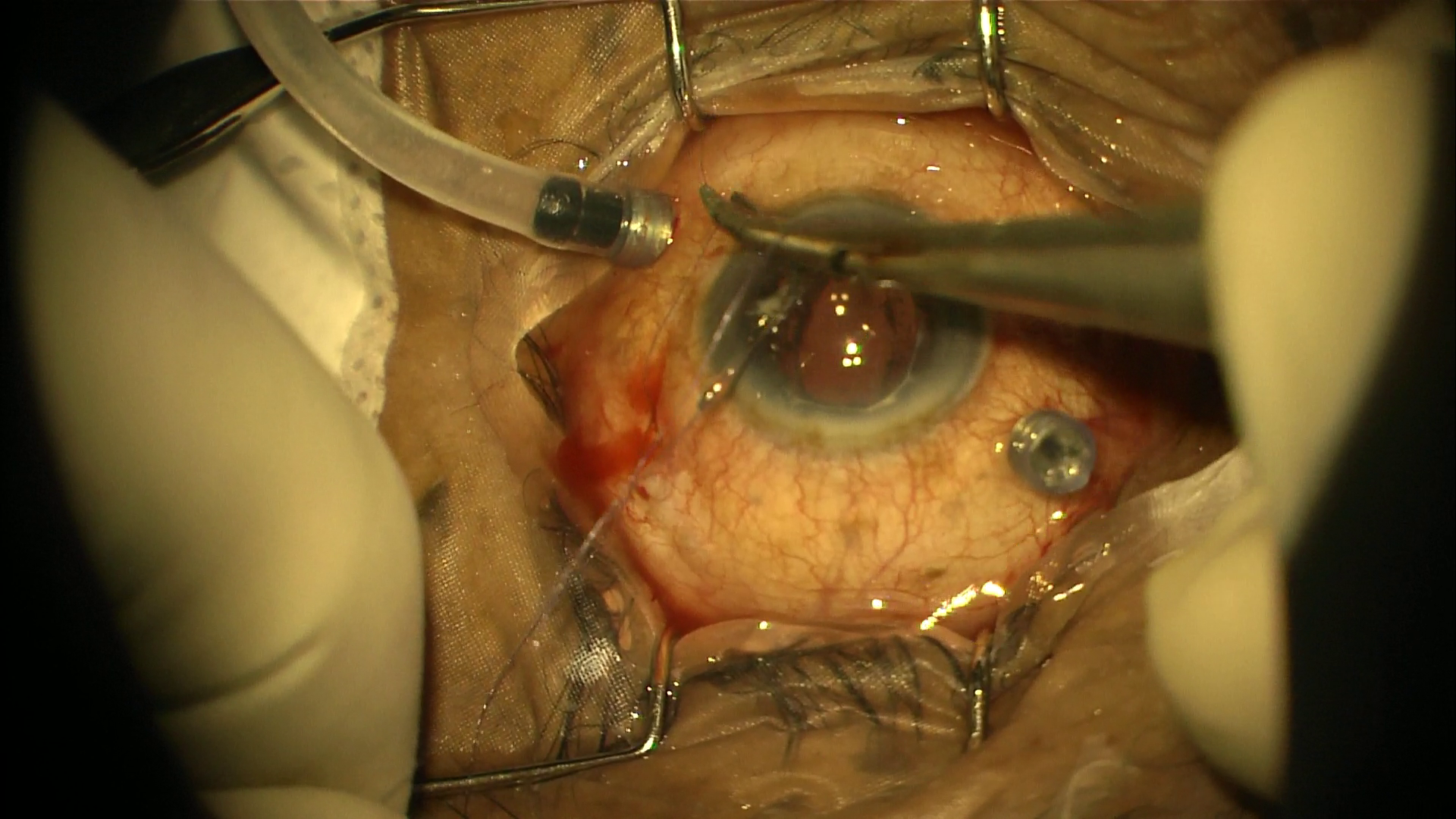
pattern and extent are tailored to the diagnosis.Watch Dr Gupta explain retinal laserVitrectomy in progressA real intraoperative view of small-gauge vitreoretinal
surgery. The surgical approach is selected according to the
retinal condition, its severity and the needs of the
individual eye.
Dr Shashank Rai GuptaSenior Vitreo-Retina Surgeon · Founder Chairman, Delhi Eye
Care
Experience with perspective
Retina expertise, translated into understandable decisions.
Dr Shashank Rai Gupta leads the Retina Excellence Centre at Delhi
Eye Care. His work spans medical retina, diabetic retinal disease,
macular disorders, uveitis and vitreoretinal surgery. He
previously served as Senior Consultant and Head of Vitreo-Retina &
Uveitis at Vasan Eye Care Hospitals, New Delhi, and as Consultant
at Venu Eye Institute & Research Centre.
17 yearsdevoted to retina practice
7,468retinal procedures and surgeries
28,700intravitreal injections and retinal laser procedures
Experience figures supplied by Delhi Eye Care and updated
September 2026.
Patient experiences are individual; outcomes and recovery vary
according to the condition and treatment.
Questions patients often ask
Clear answers before the first visit.
Are flashes and floaters always dangerous?
Not always, but a sudden onset or marked increase can accompany
a retinal tear or detachment. A dilated retinal examination
helps determine the cause.
Is retinal detachment an emergency?
A suspected retinal detachment needs prompt specialist
assessment. Timing and treatment depend on its location, extent,
symptoms and the condition of the macula.
Does every person with diabetes need a retinal examination?
Diabetes can affect the retina before vision changes. The timing
and frequency of examination depend on diabetes type, duration,
pregnancy, retinal findings and other health factors.
What is an OCT scan?
OCT is a non-contact scan that produces cross-sectional images
of the retina. It helps assess the macula, retinal layers and
fluid, and compare change over time.
Does every retinal condition require surgery?
No. Some conditions need observation, medical treatment, laser
or injections. Surgery is recommended only when its expected
benefit justifies the procedure and timing.
Can I request a second opinion using existing reports?
Yes. Previous prescriptions, OCT scans, angiography, retinal
photographs and surgical advice can help the specialist
understand your history. A final plan may still require
examination.
Your next step
Concerned about your retina—or already have reports?
Speak with the Delhi Eye Care team to arrange a retina consultation
or specialist report review.
Send only relevant eye reports and remove unrelated identity or
financial documents. WhatsApp is a third-party service and is
not for emergencies; a report review does not replace an
examination.